Our Health Library information does not replace the advice of a doctor. Please be advised that this information is made available to assist our patients to learn more about their health. Our providers may not see and/or treat all topics found herein.
Chest X-Ray
Test Overview
A chest X-ray is a picture of the chest that shows your heart, lungs, airway, blood vessels, and lymph nodes. A chest X-ray also shows the bones of your spine and chest, including your breastbone, your ribs, your collarbone, and the upper part of your spine.
A chest X-ray can help find some problems with the organs and structures inside the chest. Usually two pictures are taken, one from the back of the chest and another from the side. In an emergency when only one X-ray picture is taken, a front view is usually done. Doctors may not always get the information they need from a chest X-ray to find the cause of a problem. If the results from a chest X-ray are not normal or do not give enough information about the chest problem, more specific X-rays or other tests may be done, such as a computed tomography (CT) scan, an ultrasound, an echocardiogram, or an MRI scan.
Why It Is Done
A chest X-ray is done to:
- Help find the cause of common symptoms such as a cough, shortness of breath, or chest pain.
- Find lung conditions—such as pneumonia, lung cancer, chronic obstructive pulmonary disease (COPD), collapsed lung (pneumothorax), or cystic fibrosis—and monitor treatment for these conditions.
- Find some heart problems, such as an enlarged heart, heart failure, and problems causing fluid in the lungs (pulmonary edema), and to monitor treatment for these conditions.
- Look for problems from a chest injury, such as rib fractures or lung damage.
- Find foreign objects, such as coins or other small pieces of metal, in the tube to the stomach (esophagus), the airway, or the lungs. A chest X-ray may not be able to see food, nuts, or wood fibers.
- See if a tube, catheter, or other medical device has been placed in the proper position in an airway, the heart, blood vessels of the chest, or the stomach.
How To Prepare
In general, there's nothing you have to do before this test, unless your doctor tells you to.
How It Is Done
You will need to take off jewelry that might be in the way of the X-ray picture. You may need to take off all or most of your clothes above the waist. (You may be allowed to keep on your underwear if it does not get in the way of the test.) You will be given a gown to wear during the test.
Two X-ray views of the chest are usually taken. One view is taken from the back. The other view is taken from the side of the body. But other views may be needed, depending on what your doctor is looking for. In an emergency, only one picture may be taken, usually from the front.
You will probably stand with your front against an X-ray plate for the pictures. If you need to sit or lie down, someone will help you get into the correct position.
You will need to hold very still during the X-ray to prevent blurring of the picture. You may be asked to hold your breath for a few seconds while the X-ray picture is taken.
Most hospitals and some clinics have portable X-ray machines. If a chest X-ray is done with a portable X-ray machine at your bedside in a hospital, an X-ray technologist and nurse will help you move into the correct position. Usually only one picture from the front is taken.
How long the test takes
The test will take about 10 minutes.
Watch
How It Feels
You won't feel any pain from the X-ray itself. If you have pain from your chest problem, you may feel some discomfort if you need to hold a certain position, breathe deep, or hold your breath while the X-ray is done.
Risks
There is always a slight chance of damage to cells or tissue from radiation, including the low levels of radiation used for this test. But the chance of damage from the X-rays is extremely low. It is not a reason to avoid the test.
If you need an X-ray while you are pregnant, a lead apron will be put over your belly to protect the baby from exposure to radiation from the X-rays. The chance of harm is usually very low compared with the benefits of the test.
Results
In an emergency, the results of a chest X-ray can be available within a few minutes for review by your doctor. If it is not an emergency, results are usually ready in 1 or 2 days.
Normal:
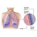
- The lungs look normal in size and shape, and the lung tissue looks normal. No growths or other masses can be seen within the lungs. The spaces surrounding the lungs (pleural spaces) also look normal.
- The heart looks normal in size and shape, and the heart tissue looks normal. The blood vessels leading to and from the heart also are normal in size, shape, and appearance.
- The bones including the spine and ribs look normal.
- The diaphragm looks normal in shape and location.
- No abnormal collection of fluid or air is seen, and no foreign objects are seen.
- All tubes, catheters, or other medical devices are in their correct positions in the chest.
Abnormal:
- An infection, such as pneumonia or tuberculosis, is present.
- Problems such as a tumor, injury, or a condition such as edema from heart failure may be seen. In some cases, more X-rays or other tests may be needed to see the problem clearly.
- A problem such as an enlarged heart—which could be caused by heart damage, heart valve disease, or fluid around the heart—is seen. Or a problem of the blood vessels, such as an enlarged aorta, an aneurysm, or hardening of the arteries (atherosclerosis), is seen.
- Fluid is seen in the lungs (pulmonary edema) or around the lungs (pleural effusion), or air is seen in the spaces around a lung (pneumothorax).
- Broken bones (fractures) are seen in the rib cage, collarbone, shoulder, or spine.
- Enlarged lymph nodes are seen.
- A foreign object is seen in the esophagus, breathing tubes, or lungs.
- A tube, catheter, or other medical device looks like it has moved out of the correct position.
Related Information
Credits
Current as of: July 31, 2024
Author: Ignite Healthwise, LLC Staff
Clinical Review Board
All Ignite Healthwise, LLC education is reviewed by a team that includes physicians, nurses, advanced practitioners, registered dieticians, and other healthcare professionals.
Current as of: July 31, 2024
Author: Ignite Healthwise, LLC Staff
Clinical Review Board
All Ignite Healthwise, LLC education is reviewed by a team that includes physicians, nurses, advanced practitioners, registered dieticians, and other healthcare professionals.
This information does not replace the advice of a doctor. Ignite Healthwise, LLC disclaims any warranty or liability for your use of this information. Your use of this information means that you agree to the Terms of Use and Privacy Policy. Learn how we develop our content.
To learn more about Ignite Healthwise, LLC, visit webmdignite.com.
© 2024-2025 Ignite Healthwise, LLC.


